 | |
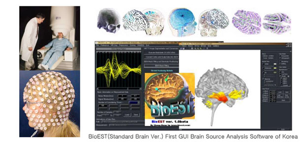
BioEST - A free software to reconstruct EEG/MEG source distribution developed by Dr. Chang-Hwan Im
1. What is BioEST™? (EEG Standard Brain Version)
BioEST™ is a general purpose brain electrical source estimation software based upon cortically distributed source model. In the title ‘BioEST’, EST represents ‘estimation’. The BioEST estimates brain electrical sources on a standard Talairach brain based on cortically distributed source model. This software would be very useful especially when there is no individual MRI T1 images or no head anatomical data. The main features of the BioEST are listed below:
(1) Generalized source reconstruction software – easily applicable to any kinds of data with just slight modifications
(2) Boundary element method (BEM) was used for forward calculation. Fast matrix solver (LAPACK routine) was adopted.
(3) Anatomical constraints are used. Linear (Wiener) inverse estimation is used for the reconstruction. Cortical meshes are segmented and tessellated from MRI T1 images.
(4) Accuracy of the software was verified extensively using several experimental data.
(5) FOCUSS (Focal underdetermined system solution) algorithm can be used optionally.
(6) Standard MNI (Montreal Neuroscience Institute) Brain was used for the model. (http://www.mrc-cbu.cam.ac.uk/Imaging/Common/mnispace.shtml)
2. BioEST™ ver. 1.5beta (trial version) User Manual
pdf version of manual
3. Download of BioEST™
If you want to try this software, please send me an E-mail containing your name, affiliation, and your E-mail address. You will be automatically registered in the user group of BioEST and can get information on latest version of BioEST. If you find any bugs in the current version, please do not hesitate to let me know. Thank you for using this software!
You can send an E-mail to ich@hanyang.ac.kr.
The download site is temporarily closed. Please send me an E-mail if you want to try this software.
4. Examples of Results
Download Example Data Sets (included in the program package) - interictal EEG acquired from an epilepsy patient
* Example 1 (08-16-2005) - A comparison between LORETA-key and BioEST
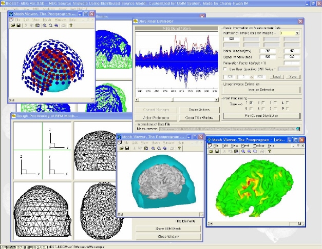
New Release!!! BioEST MEG ver. 0.5 beta
Use of Individual Brain Anatomy Extracted from Structural MRI
Neuromag Axial Gradiometer System (Customized for U of M MEG System)
E-mail: ich@hanyang.ac.kr, 2011 (c) all rights are reserved
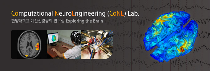
Computational
NeuroEngineering Laboratory, Hanyang University